Diagnosing MS: Referring to the Revised McDonald Criteria
Patricia K. Coyle, MD, highlights revisions to the McDonald diagnostic criteria for multiple sclerosis and emphasizes why health care professionals need to start referring to the criteria in a more standard fashion.
Episodes in this series

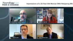
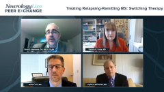
Scott D. Newsome, DO, MSCS, FAAN: Hello, and welcome to this Neurology Live® Peer Exchange titled “Strategies in Treating Relapsing/Remitting Multiple Sclerosis.” I am Dr Scott Newsome from the Johns Hopkins Multiple Sclerosis and Transverse Myelitis Center in Baltimore, Maryland. Joining me in this discussion are esteemed colleagues. First, Dr Patricia Coyle from Stony Brook Neuroscience Institute in Stony Brook, New York; Dr Robert Fox from the Cleveland Clinic in Cleveland, Ohio; and Dr Clyde Markowitz from the Multiple Sclerosis Center at Penn Medicine in Philadelphia, Pennsylvania. Today we are going to discuss a number of topics pertaining to the diagnosis and treatment of relapsing/remitting multiple sclerosis. Let’s get started on our first topic.
Pat, can you tell us a bit about the revised McDonald criteria? Specifically, what key changes in the 2017 criteria are helping clinicians in the clinic make diagnoses sooner and helping in terms of the treatment related to multiple sclerosis?
Patricia K. Coyle, MD: To start out, we have just 1 diagnostic criterion set forth in MS that is being used, and it was most recently revised in 2017. The revised McDonald criteria is the third evolution; we should be using this in the United States routinely, and we are not. We need to push to have this used. The latest revision focused in particular on misdiagnoses, so they indicated that these criteria should be applied to very crisp, clinically isolated syndromes like optic neuritis, transverse myelitis, isolated brain stem, or cerebellar syndrome. Not headache, not foggy brain, not muscle and joint pain, and not paresthesias.
Of course, they had formal definition for primary progressive MS [multiple sclerosis]. They said that we could count symptomatic lesions now. They added cortical lesions to juxtacortical. They also clarified that MRI lesions had to be at least 3 mm. You cannot count those smaller than that. If you called it periventricular, then it had to abut the ventricles. People were miscalling ventricular lesions that were not. The juxtacortical lesions had to abut the cortex. They clarified that. They also brought spinal fluid back, and they elevated CSF [cerebrospinal fluid]–specific oligoclonal bands. They did not require it, but they strongly urged using CSF in more unusual populations if there was any uncertainty at all if you were not going with an absolutely classic case. They also did not mandate spinal cord imaging, but that would be helpful as well.
They indicated that, although this is a clinical diagnosis, for the diagnostic criteria, you are obligated to create a differential diagnosis to rule it out so that MS is left as the most likely diagnosis. To me, that is selected blood work as well as imaging of the brain and the entire spinal cord: the central nervous system. We include OCT [optical coherence tomography] as part of looking at the central nervous system and interrogating that. We include spinal fluid testing in all patients.
I will add something that may be a bit controversial: In relapsing MS, we are checking aquaporin-4 and NMO [neuromyelitis optica]–IgG in the blood of all our patients now.
Scott D. Newsome, DO, MSCS, FAAN: That is a great point. We are doing more of that as well because as we learn about the full spectrum of radiological findings in MOG [myelin oligodendrocyte glycoprotein] and NMOSD [neuromyelitis optica spectrum disorder], we are finding that some of the patients who we thought had MS in years past actually fall under NMOSD and MOGAD [myelin oligodendrocyte glycoprotein antibody disease].
Thank you for watching this Neurology Live® Peer Exchange. If you enjoyed the content, and I hope you did, please subscribe to our e-newsletters to receive upcoming Peer Exchanges and other great content right in your in-box.
Transcript Edited for Clarity
Newsletter
Keep your finger on the pulse of neurology—subscribe to NeurologyLive for expert interviews, new data, and breakthrough treatment updates.

